CHRISTUS Children’s Uses 3D Printing to Guide Surgeons Through Complex Heart and Orthopedic Cases
CHRISTUS Children’s is widely recognized across the region for its highly specialized services, including its ability to perform complex surgeries to address congenital heart defects and orthopedic conditions. As part of its comprehensive program, the hospital also leads the way in advanced imaging and pre-surgical three-dimensional (3D) modeling, which helps ensure the best possible outcomes for patients.
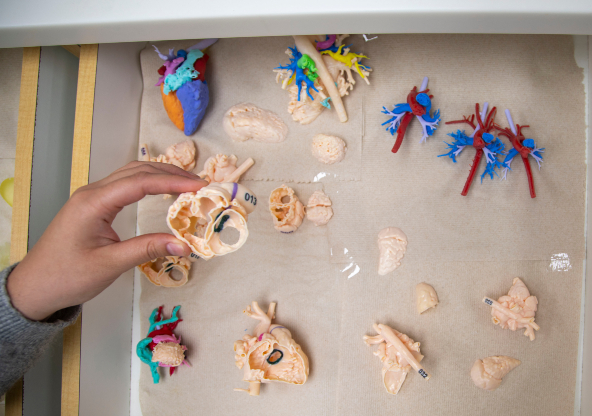
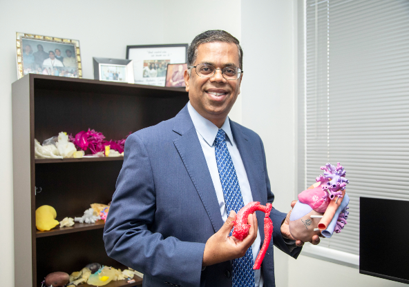
At the forefront of this effort is Dr. Ravi Ashwath, Division Chief of Pediatric Cardiology at CHRISTUS Children’s, who leads the 3D printing and modeling program. Under his leadership, the program has grown into one of only a handful of its kind in the U.S., offering 3D modeling for complex conditions to help surgeons gain a deeper understanding of each child’s unique anatomy. When printed, these 3D models provide an exact reproduction surgeons can hold in their hands, allowing them to physically prepare ahead of a procedure. This technology delivers the clearest possible picture of a patient’s condition and supports the creation of a customized surgical plan best suited to each patient.
The imaging program has been especially beneficial for surgeons treating complex congenital heart conditions. In these cases, the center creates 3D models prior to surgery, giving specialists the most detailed view possible of a patient’s condition. This allows them to plan more precisely and prepare more effectively for the operation, often leading to shorter operating times and fewer complications.
To create advanced visualizations of cardiac anatomy, we begin with raw imaging data obtained from computed tomography (CT) or magnetic resonance angiography (MRA) scans. These scans provide high-resolution, two-dimensional (2D) cross-sectional images of the patient’s heart. This raw data is then segmented by highly trained imaging specialists using specialized software. Segmentation involves isolating relevant anatomical structures—such as chambers, vessels and valves—from surrounding tissues. This process is meticulous and can take several hours depending on the complexity of the anatomy and the quality of the source images.
Once segmentation is complete, the data is converted into a standard tessellation language (STL) format, which is compatible with 3D modeling platforms. The STL file can then be used in two key ways:
- 3D Printing:
The STL model can be printed using medical-grade 3D printers to produce a physical replica of the patient’s heart. This tangible model allows clinicians and surgeons to visualize and manipulate the anatomy, aiding in pre-surgical planning and education. - Augmented Reality (AR) and Virtual Reality (VR):
The same STL model can be imported into AR/VR platforms, enabling immersive visualization. Using AR headsets, clinicians can interact with the virtual heart model in three dimensions, rotate it and examine internal structures from any angle. This enhances spatial understanding and supports collaborative decision-making.
At CHRISTUS Children’s, surgeons are leveraging technologies like 3D printing and augmented/virtual reality (AR/VR) to revolutionize the way complex congenital heart conditions are understood and treated. These tools allow clinicians to explore the intricate spatial relationships within and around the heart in ways that traditional imaging simply cannot match. Because the human body is inherently three-dimensional, this approach provides the most accurate and intuitive representation of a patient’s unique anatomy.
This technology is particularly valuable in the treatment of congenital heart disease, where no two cases are exactly alike. By studying a patient-specific 3D model, surgeons gain a deeper understanding of the defect and can plan each step of the procedure with precision. This level of preparation has been shown to reduce overall surgical time, minimize the duration a child spends on heart-lung bypass machine and lower the risk of intraoperative complications.
For patients with especially complex cardiac anatomy, a 3D-printed replica of the heart can be physically opened and examined mirroring the surgical approach. This allows the surgical team to rehearse the procedure in advance, ensuring that every decision is deliberate and well-informed.
Beyond surgical planning, these models serve as powerful tools for patient and family education. They help translate complex medical information into tangible, understandable visuals, fostering better communication and shared decision-making.
“These models have transformed how we communicate with families,” said Dr. Ashwath. “With a precise 3D replica of their child’s heart, we can clearly show the anatomy and explain the surgical plan in a way that’s both visual and easy to understand. It’s a powerful tool that brings clarity and reassurance during a difficult time. Instead of relying solely on flat images or abstract descriptions, we can now present a precise, three-dimensional replica of their child’s heart. It’s an incredibly useful tool to help families grasp the magnitude of the surgery.”
While the Advanced Cardiac Non-Invasive Imaging and 3D+ Program at CHRISTUS Children’s focuses primarily on complex congenital heart cases, the cardiology team can also use 3D modeling for nonstandard orthopedic cases, complex neurosurgical procedures and craniofacial plastic surgery.
“The 3D models are valuable tools for our orthopedic surgeons, particularly when treating children with complex hip and spinal deformities,” said Dr. Shawn Funk, a pediatric orthopedic surgeon and medical director of the Scoliosis and Spinal Deformity Program at CHRISTUS Children’s. “Being able to see the geospatial relationship ahead of surgery is a game changer and it helps us plan surgery based on each patient’s unique anatomy.”
Looking ahead, the team is working to advance technology and expand its capabilities across additional clinical areas. CHRISTUS Children’s is also becoming one of the few programs in the country to offer highly specialized imaging technology for the fetal heart, allowing surgeons to prepare for critical, life-saving surgeries even before birth.
This program reflects our commitment to innovation and excellence in pediatric cardiac care,” said Dr. Ashwath. “By providing surgeons with detailed, patient-specific models—even prenatally—we’re enhancing surgical precision and improving outcomes for our youngest patients.” “We’re proud to offer this service, which benefits our surgeons in their work and ultimately, helps us provide the best possible care and treatment for our patients.”
